ChestView
AI solution for critical chest pathology
ChestView offers radiologists and emergency department doctors an instant, automated second opinion on chest X-rays, seamlessly integrated into the reading workflow.


ChestView assists in identifying critical chest pathologies, such as pneumothorax, consolidation, nodule, mediastinal mass, and pleural effusion, enhancing the detection of urgent findings and early cancer indicators.
Experience the power of ChestView
Indication
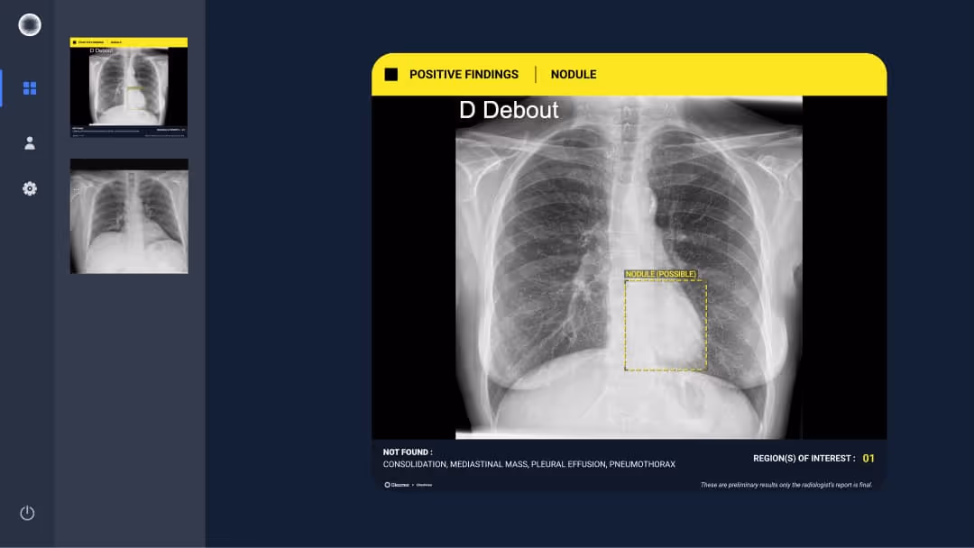
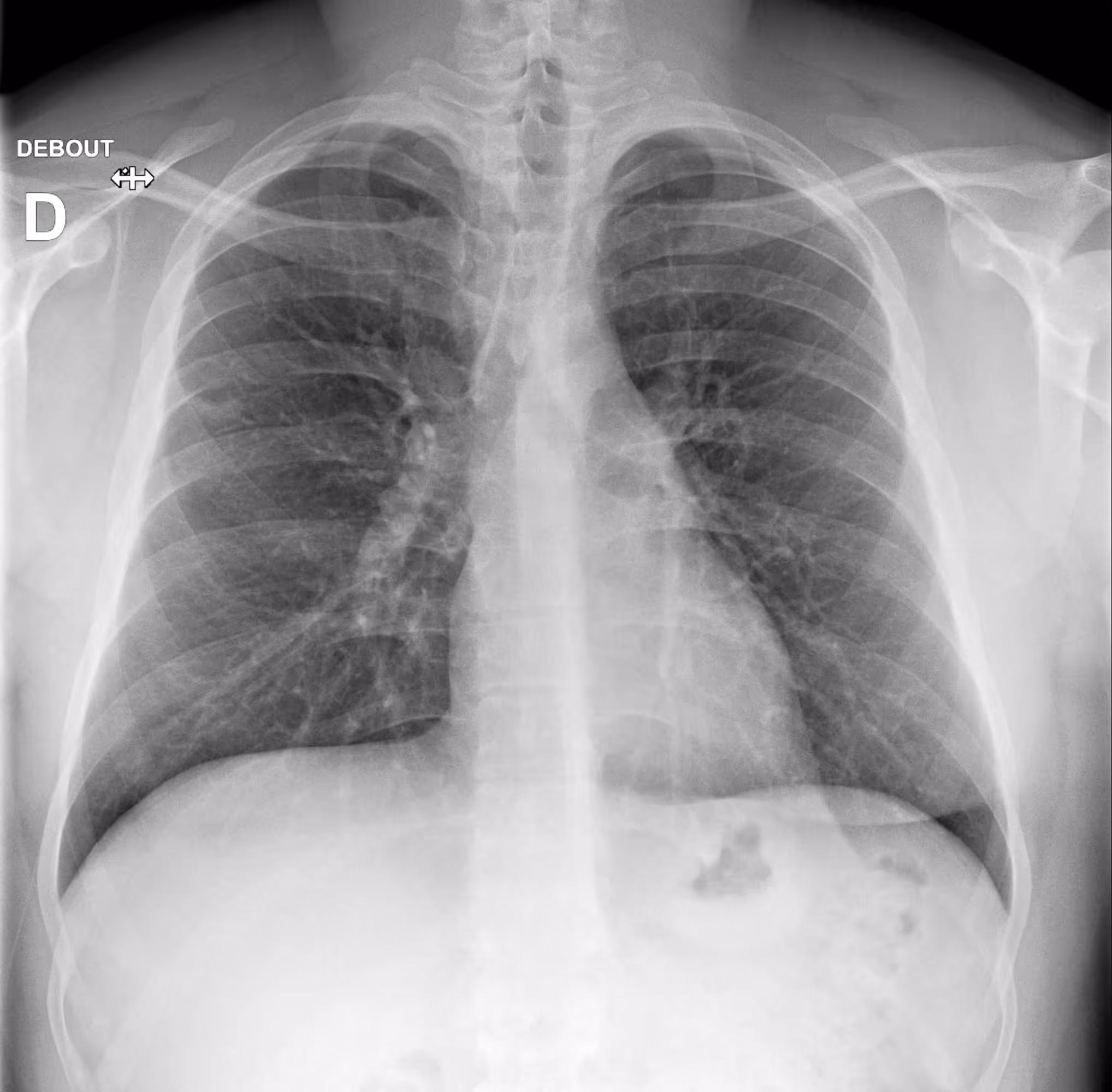
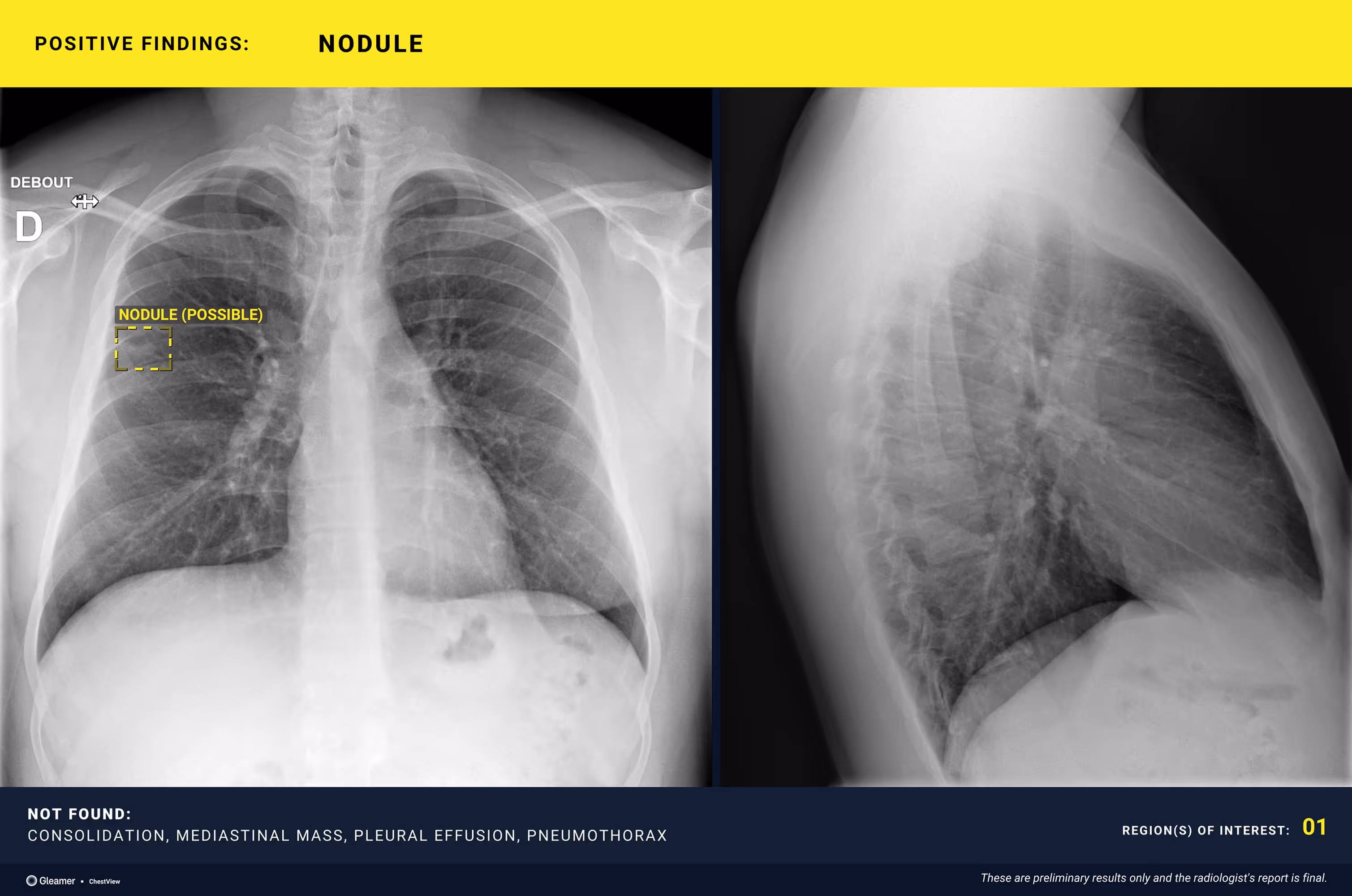
A 26-year-old male smoker with persistent cough.
Results
ChestView detected a suspicious nodule, confirmed by CT.
Indication
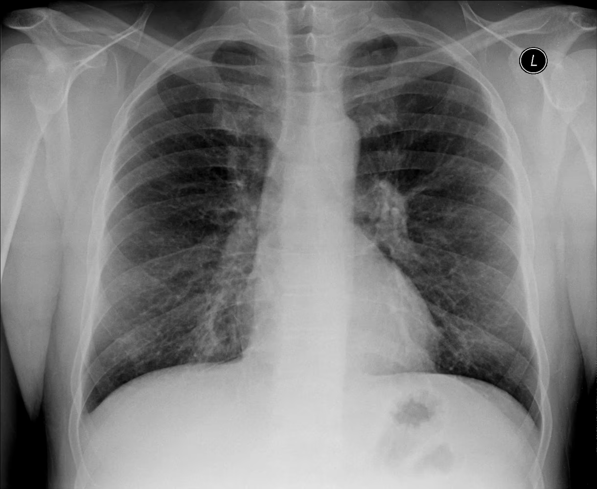
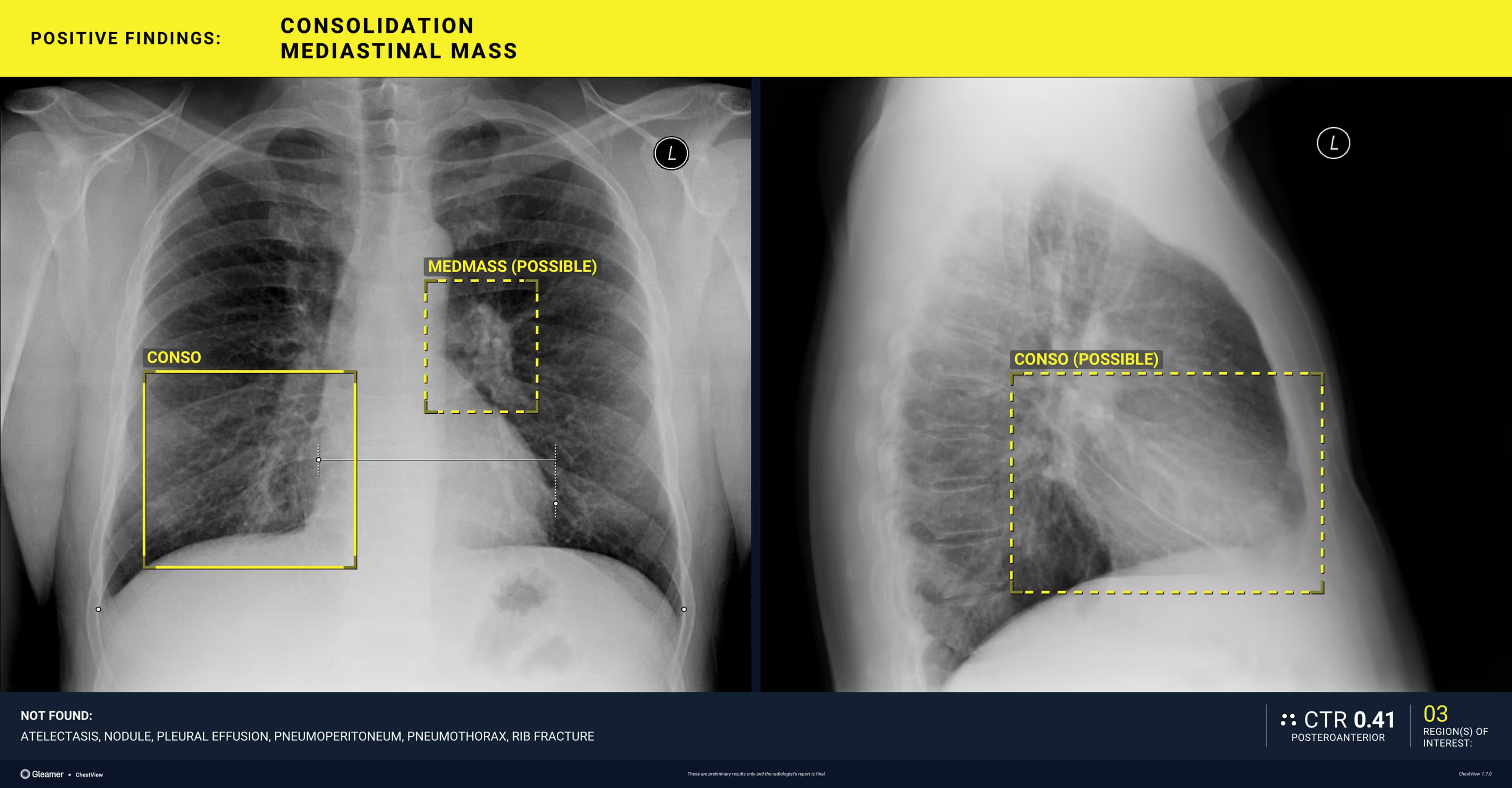
A 54-year-old male with respiratory difficulty.
Results
ChestView detected an infectious region in the right lower lobe and incidentally identified left hilar lymphadenopathy
Indication
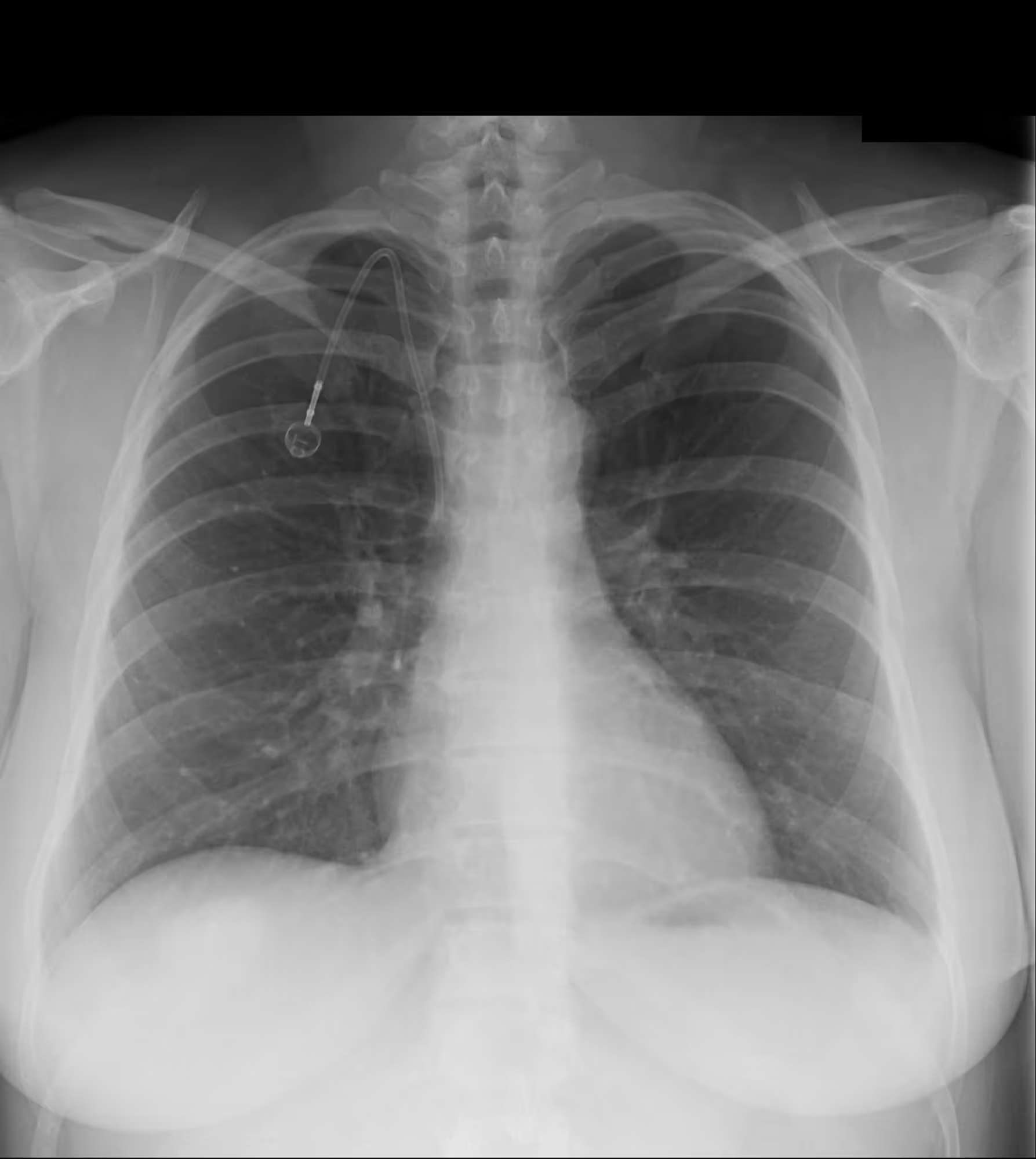
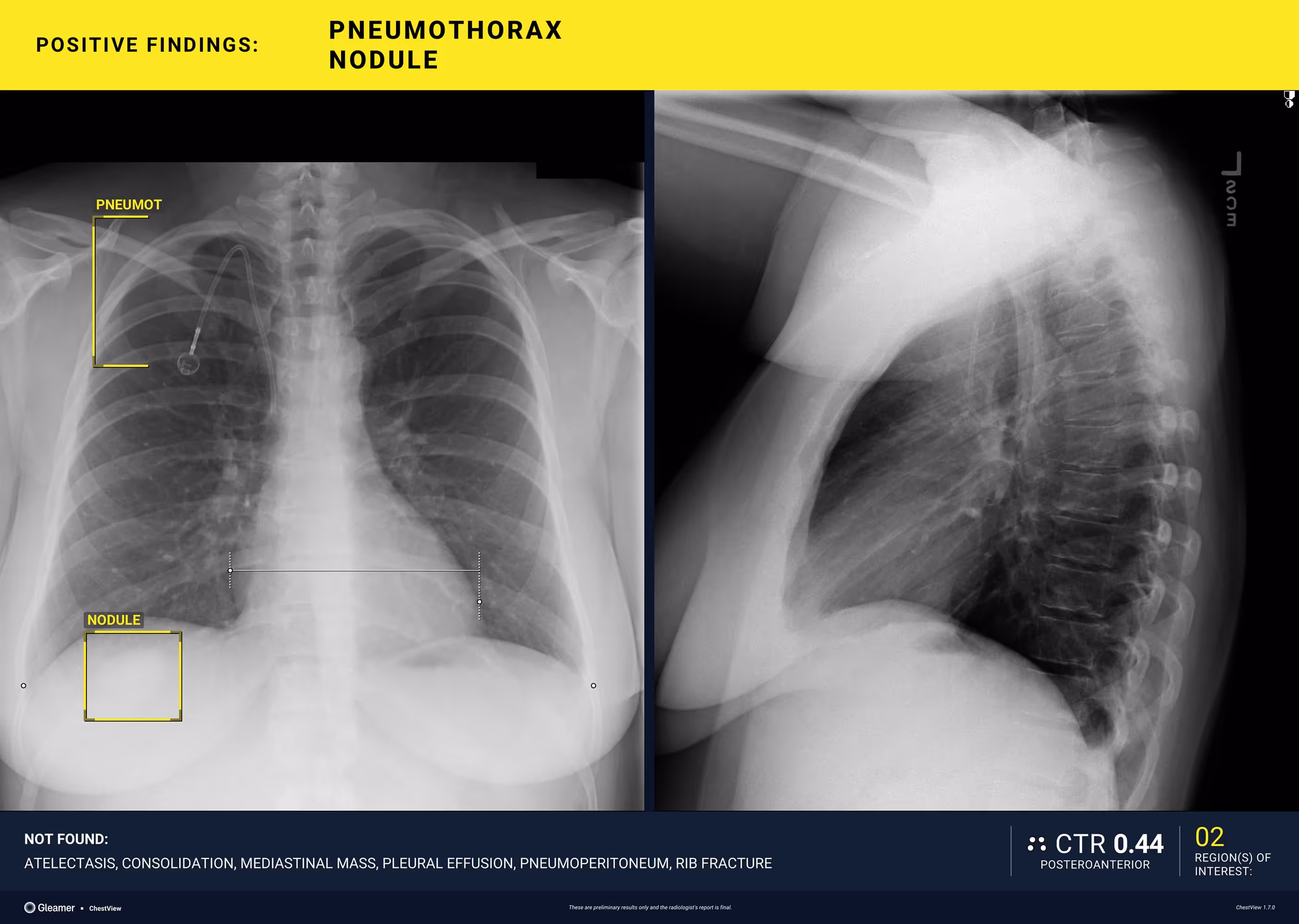
A 40-year-old female with a history of colon cancer presents for a control radiograph following biopsy of a right lower-lobe pulmonary nodule.
Results
ChestView detected a pneumothorax post-biopsy.
Indication
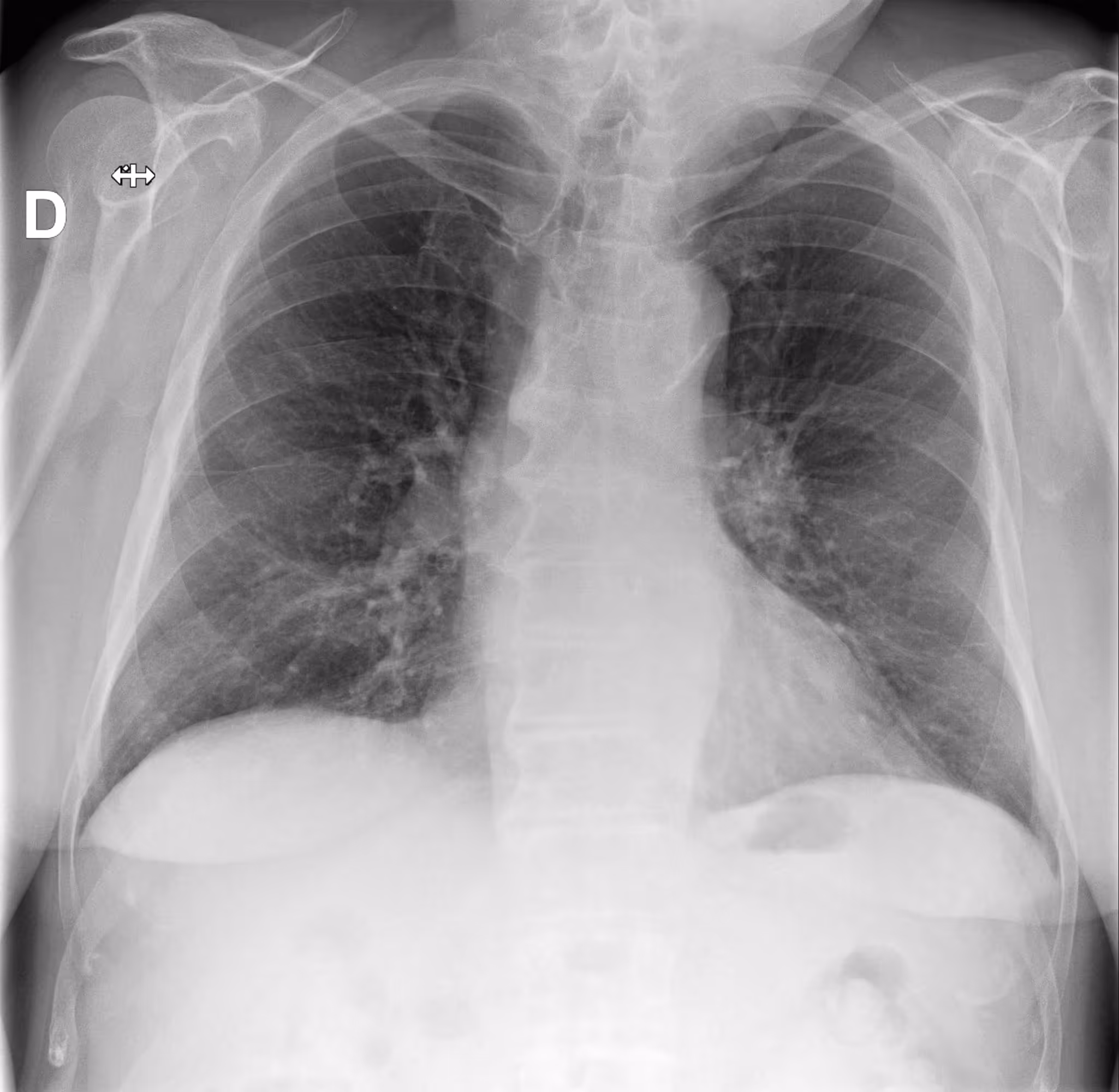
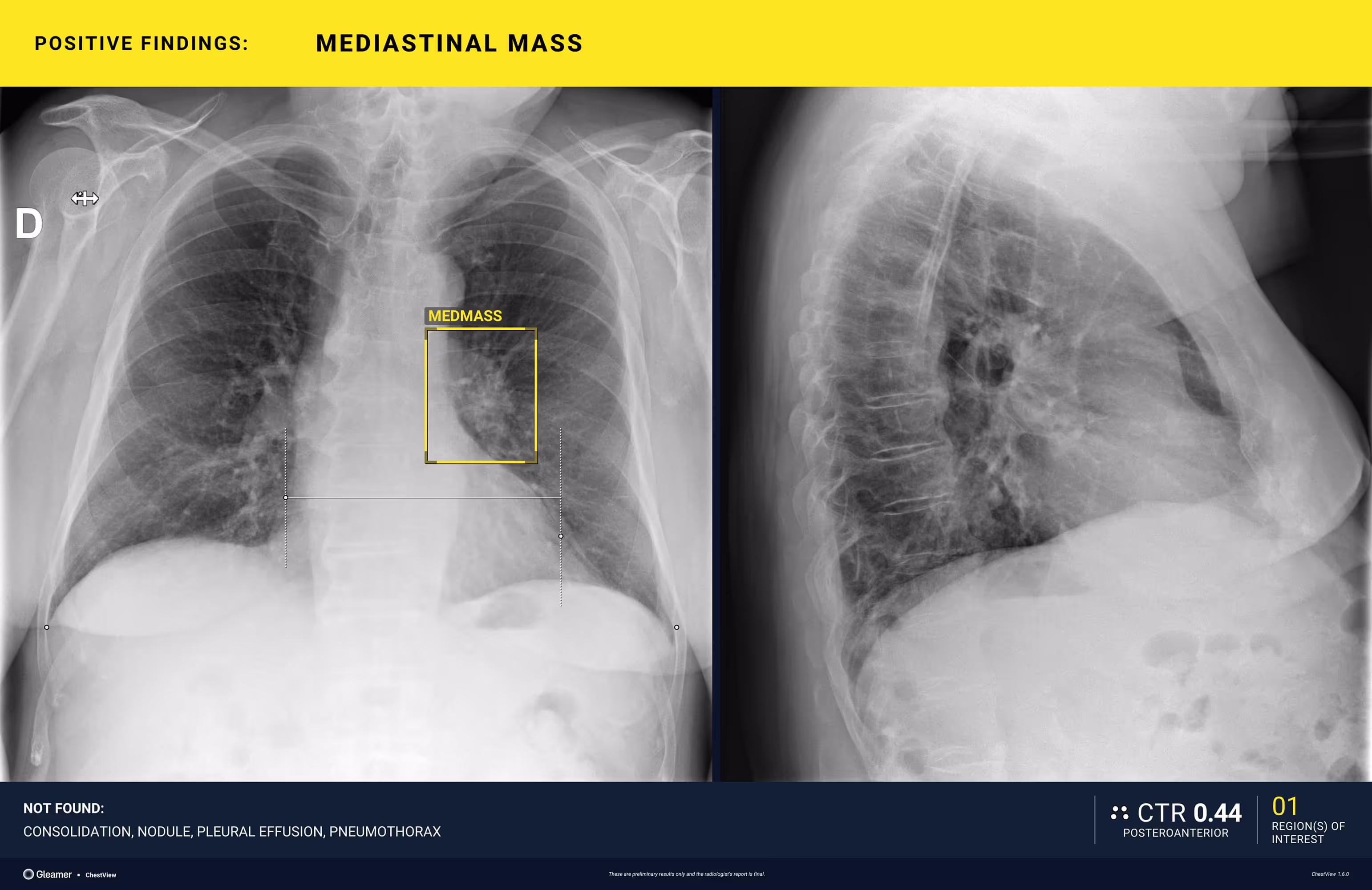
An 80-year-old male with a previously normal chest X-ray presents for CT evaluation 9 months later, which reveals findings suggestive of lung cancer.
Results
ChestView detected the mediastinal mass on the initial X-ray.
Specific features
Discover more about ChestView
Automatically measures the Cardiothoracic Ratio
ChestView automatically measures the Cardiothoracic Ratio (CTR) on posteroanterior (PA) chest X-rays for patients over 15 years old. This measurement supports the detection of cardiomegaly, adding a valuable clinical indicator to the report and reinforcing ChestView’s role in identifying major thoracic findings.
Co-developed with AP-HP and grounded on a robust database partly cross-referenced with CT-scan, it is now widely used in private and public facilities worldwide.
Clinical Studies
ChestView
Efficacy of a deep learning-based software for chest X-ray analysis in an emergency department.
Sathiyamurthy Selvam, Olivier Peyrony, Arben Elezi, Adelia Braganca, Anne-Marie Zagdanski, Lucie Biard, Jessica Assouline, Guillaume Chassagnon, Guillaume Mulier, Constance de Margerie-Mellon
ChestView
Using AI to improve radiologist performance in detecting abnormalities on chest radiographs
Bennani, Souhail; Regnard, Nor-Eddine; Ventre, Jeanne; Lassalle, Louis; Nguyen, Toan; Ducarouge, Alexis; Dargent, Lucas; Guillo, Enora; Gouhier, Elodie; Zaimi, Sophie-Hélène; Canniff, Emma; Malandrin, Cécile; Khafagy, Philippe; Koulakian, Hasmik; Revel, Marie-Pierre; Chassagnon, Guillaume
ChestView
Learning from the machine: AI assistance is not an effective learning tool for resident education in chest x-ray interpretation
Chassagnon G, Billet N, Rutten C, Toussaint T, Cassius de Linval Q, Collin M et al.
Boost your workflow with the power of advanced integrations
Designed with radiologists, our workflow integrations blend effortlessly into daily routines, enhancing speed, clarity, and confidence at every step.
Reporting has never been this fast
AutoReport automatically generates patient reports based on its findings. It fills in the indication, technique, and delivers smart, clinically relevant impressions, saving time while ensuring consistency and quality.


AI-powered Worklist
Worklist now highlights AI results, findings, and automatically prioritizes urgent patient cases.
Shadow Mode
AI results appear on native images, where radiologists can review, accept, or reject, all within their workflow.

Part of Gleamer Copilot
Gleamer Copilot is the all-in-one AI platform that supports radiologists from image to report. It combines powerful detection tools, smart measurements, and structured reporting to boost accuracy and efficiency, all seamlessly integrated into your workflow.
¹Relative improvement of sensitivity when readers used ChestView for all pathologies: +50.0% for pneumothorax, from 52.4% to 78.6%. (Bennani et al., Radiology, 2023)
²Missed pneumothorax decreased by 55% with AI assistance. Relative reduction in lesion wise FNR ; 47.6% to 21.4%. (Bennani et al., Radiology, 2023)
³Decreased reading time by 38% for radiographs with no abnormalities. From 68 seconds without AI to 42 seconds with AI. (Bennani et al., Radiology, 2023)
For the latest regulatory information, refer to: https://www.gleamer.ai/privacy-policy