Pixyl.Neuro.BV
AI solution for Neurodegenerative diseases
Pixyl.Neuro BV gives radiologists an AI-powered second reader for brain MRI. It automatically segments brain structures on 3D T1-weighted gradient echo sequences, computes volumes and compares results to normative references, highlighting any abnormalities through a clear structured report right in your workflow.


Quantify and track brain structures volumes
Pixyl.Neuro.BV supports differential and early diagnosis of neurodegenerative diseases. It segments the whole brain, gray/white matter, ventricles, hippocampi, cortical lobes and other key regions, computes volumes and tracks change over time, so you can standardize reporting, support earlier detection of abnormal atrophies and monitor progression with confidence.
See Pixyl.Neuro.BV in Action
Indication
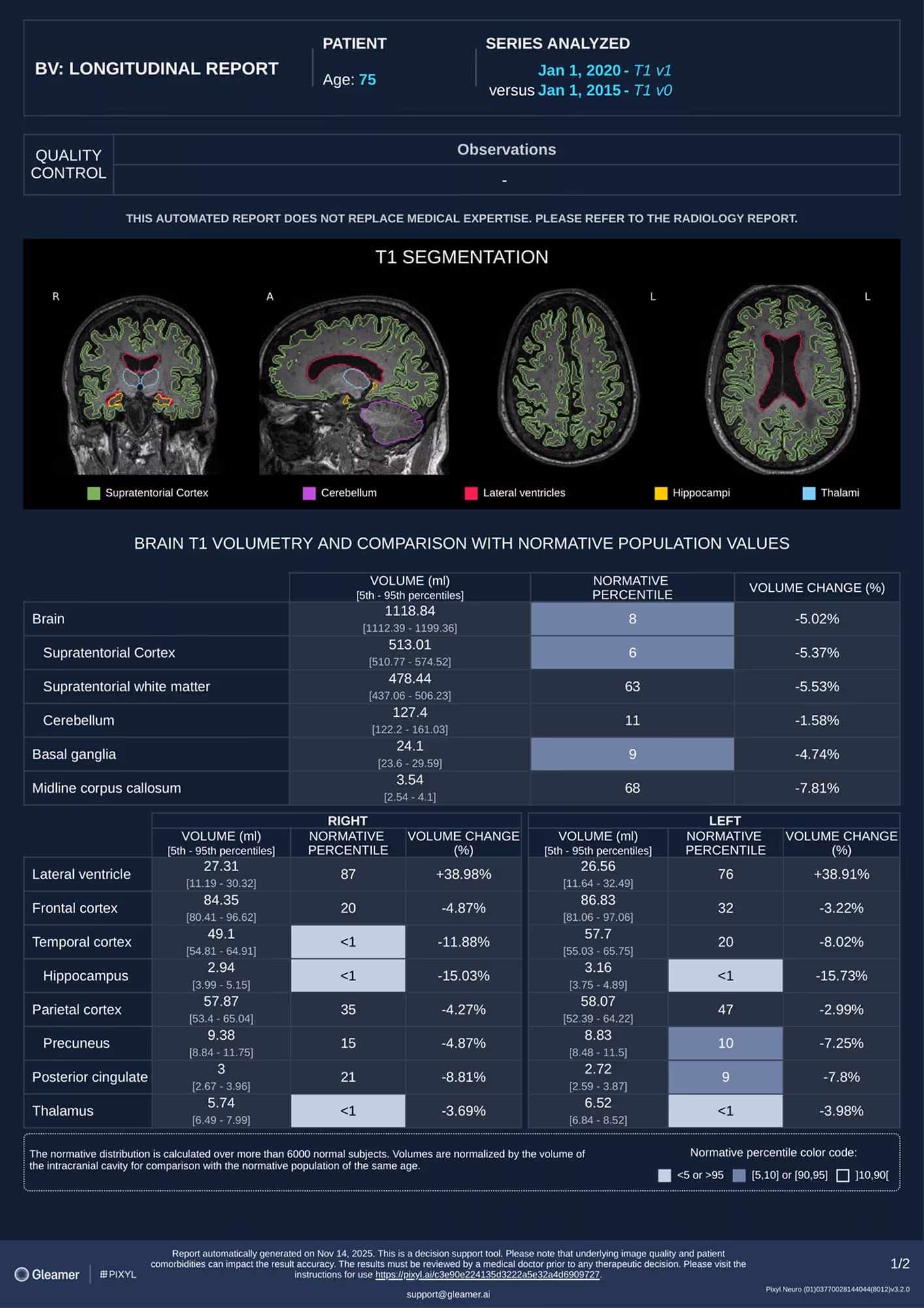
A 75-year-old patient presents with a strong dementia.
Results
Pixyl.Neuro.BV reported pathological right temporal atrophy progressing faster than normal aging, supporting a diagnosis of Alzheimer’s disease.
For Adults
Pixyl.Neuro.BV is intended to be use on brain MRIs of patients aged between 18 and 90.
Compatible with routine MRI
Works with 3D T1 Gradient Echo (single or multiple timepoints)
Specific features
Discover more about Pixyl.Neuro.BV
Atrophy quantification with respect to normative population
Provides percentiles compared to the healthy control population of the same age for each individual quantified brain structure. Abnormalities are flagged when values are out of typical range.
Specific patterns of atrophy highlighted
Asymmetries and other specific patterns of atrophy identified at-a-glance with a visual display (glass-brain, spider plots, plots with normative values) to support differential diagnosis.
Longitudinal change
Computes per-region change between timepoints and highlights volume evolution to support follow-up decisions and differential diagnosis.
Precision you can measure.
Boost your workflow with the power of advanced integrations
Designed with radiologists, our workflow integrations blend effortlessly into daily routines, enhancing speed, clarity, and confidence at every step.
Reporting has never been this fast
AutoReport automatically generates patient reports based on its findings. It fills in the indication, technique, and delivers smart, clinically relevant impressions, saving time while ensuring consistency and quality.


AI-powered Worklist
Worklist now highlights AI results, findings, and automatically prioritizes urgent patient cases.
Shadow Mode
AI results appear on native images, where radiologists can review, accept, or reject, all within their workflow.
