Pixyl.Neuro.MS
AI solution for multiple sclerosis on brain MRI
Pixyl.Neuro MS gives radiologists an AI-powered second reader for MS follow-up. It automatically detects, categorizes and quantifies white-matter lesions on 3D FLAIR, highlights new and enlarging lesions over time, and delivers segmented series and a clear structured report directly in your workflow.


Detect and characterize MS lesions
Pixyl.Neuro MS focuses on consistent lesion detection and longitudinal comparison. It measures lesion count, volume, and location, compares current and prior studies, and flags activity, supporting confident reporting and timely therapeutic decisions.
See Pixyl.Neuro.MS in Action
Indication
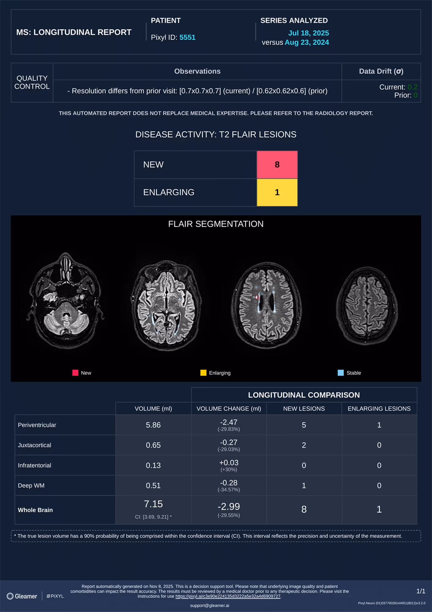
A 38-year-old male undergoing follow-up for multiple sclerosis.
Results
Pixyl.Neuro.MS reported eight additional lesions indicating continuous disease activity.
Specific features
Discover more about Pixyl.Neuro.MS
Robust activity detection
Lesions categorization: stable, enlarging, new. Colored-segmentation per category: activity between visits is highlighted allowing faster and consistent follow-up.
Accurate lesion quantification
Automatic segmentation and metrics: lesions count and volume per brain regions of interest (periventricular, juxtacortical, infratentorial, deep WM).
Regional breakdown
Reports typical MS locations (e.g., periventricular, juxtacortical, infratentorial) to support standardized reads.
Eased comparison
The prior series robustly co-registered with the current segmented series is delivered allowing series synchronization and eased comparison.
Proven impact in MS reads
Boost your workflow with the power of advanced integrations
Designed with radiologists, our workflow integrations blend effortlessly into daily routines, enhancing speed, clarity, and confidence at every step.
Reporting has never been this fast
AutoReport automatically generates patient reports based on its findings. It fills in the indication, technique, and delivers smart, clinically relevant impressions, saving time while ensuring consistency and quality.


AI-powered Worklist
Worklist now highlights AI results, findings, and automatically prioritizes urgent patient cases.
Shadow Mode
AI results appear on native images, where radiologists can review, accept, or reject, all within their workflow.

Part of Gleamer Copilot
Gleamer Copilot is the all-in-one AI platform that supports radiologists from image to report. It combines powerful detection tools, smart measurements, and structured reporting to boost accuracy and efficiency, all seamlessly integrated into your workflow.
¹All listed data are patient-wise and based on the results of an internal study.
For the latest regulatory information, refer to: https://www.gleamer.ai/privacy-policy